Having spent 3 months in the radiotherapy physics department, I finally got the opportunity today to spend some time with the radiographers, scanning, planning and treating patients. This blog post will hopefully illustrate my understanding of the pathway that patients go through for their radiotherapy treatment as well as some reflections I have had over the last few months.
Immobilisation
Immobilisation is necessary in radiotherapy to ensure that the treatment position is replicable from planning to treatment fraction to fraction. Immobilisation is most effective when the outer contour of the body matches the internal structure for example in head and neck treatments. In lower body treatments such as prostate or cervix, the internal anatomy is a bit squishier and less related to the outside anatomy and so different techniques such as bladder filling and rectal voiding are used to increase repeatibility.
The below photo is of a head and neck mask. This shell is created using a thermoplastic warmed in a water bath until soft and pliable. The patient lies on the treatment couch and the mask is applied over their face and attached to the couch with screws at the side. It hardens as it cools and forms a shell that will be used over the course of their treatment. Some centres make a mask for all their radiographers when they join the Trust to increase their empathy for the patient experience which I think is a brilliant idea!

Today we had an elderly patient for palliative care with no immobilisation specified by the referring consultant. After consulting with the superintendent radiographer, it was determined that immobilisation was definitely necessary considering the position of her tumour and its relation to her bony anatomy. However she refused to have a head and neck mask made, opting instead for a chin strap. This is a less reliable way of setting her up for treatment but the pros and cons were described to her and at the end of the day we have to respect the patient’s choice. It was really inspiring to watch the kindness with which she was treated by the radiographers, and in fact the kindness with which the radiographers treated all of their patients.
It’s very easy to forget in our Physics ivory tower (or rather windowless basement!) that everything we do concerns real people who are not equivalent to the perspex phantoms we do tests on! And there’s no point creating a technique or procedure that provides clinically better results if patients aren’t going to tolerate the treatment.
Planning CT
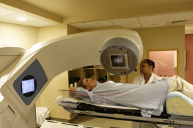
The planning CT is performed with the patient in the treatment position that they will be in during treatment. This means their immobilisation and comfort aids are used here. These include things such as the head and neck mask as discussed above but also things like knee supports, breast boards with handles that enable you to rest the arms above the head and neck cushions. All of these are designed for the comfort of the patient whilst lying on the treatment bed as well as repeatibility of the treatment position.
Patients also receive their setup tattoos during the planning CT. The lasers in the CT room are used to project points of intersection onto the patient which are marked with felt tip crosses. These crosses are then covered with radio opaque wire crosses during the CT scan so that they can be seen on the CT image. Breast patients also have any surgery scars outlined with radio opaque wire so it is visible on the CT scan as clinicians sometimes like to ensure that the entire scar is included within the radiation field jaw edges.
After the CT scan is complete, the centre of these felt tip crosses will be permanently tattooed onto the patient using ink and a needle by the radiographers. These tattoos enable the radiographers to set up a patient in the treatment room each day. They are so small I was quite impressed that the radiographers could find them so easily on the patients during treatment!

Planning
This part of the patient pathway is not seen by the patient but definitely seen by the physicists and dosimetrists!
Importing
The patient’s CT scan and information is imported into the treatment planning system. The appropriate protocol as specified by their referring clinician is attached to their CT scan and their CT scan is checked. The CT scans are checked for the date, time, number of slices and image quality to provide assurance that the correct patient scan has been imported and that all of the data has been transferred correctly (e.g. no network errors). At this point, planners will then fuse any other imaging patients have had with this CT scan. MRI images are good for soft tissue contrast but we need the electron density information from the CT scan to do our treatment planning. By fusing the image sets, we have both sets of information at our fingertips for outlining body structures and clinical target volumes as well as planning.
Contouring
After importing and any fusing, the clinicians outline the gross target volume (the tumour) and the clinical target volume (which contains a margin added in for non-imageable microscopic spread of the disease). It’s then passed back to the physics & dosimetry team for outlining of the organs at risk and planning target volume growth. The planning target volume is the clinical target volume with margins added on for geometrical uncertainties in the treatment process. These will then be approved by the clinician and it’s time to plan!
Planning
The aim of radiotherapy is to irradiate the target volume whilst minimising the dose to the healthy tissue and this is what the planning is all about. Using different sized fields, beam angles, wedges, multi-leaf collimators and segments, the planners will optimise the dose distribution such that the prescription dose to the target volume is achieved whilst minimising dose to the healthy tissue & the organs at risk in the area. In an ideal world, 100% of the dose would be given to the tumour and 0% to the healthy tissue but unfortunately this is not achievable.
The quality of the plan is assessed in a variety of ways, including looking at the dose volume histograms (i.e. how much dose is the organ receiving per percentage volume of the organ), looking at the dose distributions in different CT slices and checking the organ at risk tolerances, e.g. we want no more than 50% of the bladder to receive no more than 50 Gy. These tolerances are taken from historical data, observations & trials.
Audit
After the planning is complete and checked by an entirely independent physicist, the clinician must sign off on the plan and the plan must be checked by the radiographers. There is a weekly multidisciplinary team meeting for each treatment site in which each patient in the planning part of the patient pathway is discussed and any issues can be brought up and resolved.
On-Treatment
On the first day of a patient’s treatment, a radiographer will set aside some time to explain what to expect during treatment in terms of appointments, side effects and answering any questions they might have.
Some patients have specific preparation to undertake before every treatment. For example prostate patients will often be asked to have an enema, empty their bladder and drink 3 cups of water in the hour before their treatment. This means they have to turn up for their appointment up to an hour and a half before their allotted treatment time. Other patients have no specific preparations as such, it is very site dependent.
Imaging protocols during treatment differ between treatment sites and patients. Some sites are imaged with CBCT before every treatment and others are only imaged in the first few fractions. For example in head and neck, we expect the treatment position to be extremely reproducible each fraction due to the use of the mask however I saw a patient who was being imaged for every treatment because the differences between the set-up detailed on the set-up sheet and the set-up that was achieved on the treatment couch when aligning the lasers to the tattoos & mask crosses were greater than expected. On the bed imaging ensures the patient is in the position they are expected to be in when their treatment is delivered.
The radiographers were truly amazing in their care and patience despite a very high patient throughput. Each patient was treated with such dignity and respect (as should be the bare minimum of course!) and no one was hurried along despite the time pressures in play.
It was also interesting to see how the decisions that physics make affect the radiographers. For example when picking the isocentre for a treatment, I’ve been told to always pick a whole number of centimetres for the shift to the tattoos to make it easier for the radiographers when setting up a patient. I had suggested that surely the radiographers have enough mental maths to cope with adding 2 numbers but now having spent time with them doing patient setup I’d like to rescind all previous comments! They have more than enough to focus on without having to add non-integer numbers!
Conclusions
Physicists are vital for safe, efficacious radiotherapy treatment. However radiographers are the ones who have to deal with the consequences of the decisions that physicists make when we pick odd beam angles or strange patient shifts or decide to match or not match certain machines etc.
This day was really valuable in reminding me the importance of patient centred treatment and I’d thoroughly recommend that physicists get sent along to spend some time with radiographers in pre-treatment and on- treatment on a reasonably regular basis to be reminded of the importance of patient centred healthcare and multidisciplinary teamwork.
